Nikon Small World in Motion 2026: Winner Disqualified Over AI, Second Place Promoted
Nikon has disqualified its 2026 Small World in Motion winner over generative AI, after scientists questioned the video and a PhD student spotted a SynthID watermark in it.

- Published
- Reading time
- 5 minutes
Nikon has disqualified the winner of its 2026 Small World in Motion competition after finding the entry breached its rules on generative AI. The video appeared to show cilia beating in the airway of a child with a rare genetic disease. Scientists questioned the structures in it within days, and a PhD student found an AI watermark embedded in the footage.
That last part is the detail worth sitting with. The clip was not unmasked by a panel of judges or a forensic audit. It was unmasked, in part, because artificial intelligence now signs its own work.
The watermark
Ian Donovan, a PhD student at UT Southwestern Medical Center, spotted a SynthID watermark in the video. SynthID is the watermarking technology pioneered by Google DeepMind, designed to embed a machine-readable signature into AI-generated content invisible to a viewer, detectable by a tool that knows what to look for.
A graduate student found a machine-readable AI fingerprint inside a prize-winning scientific image. Whatever happens to this particular contest, that is the part with consequences beyond it: the provenance of a scientific image is becoming checkable by anyone who thinks to check.
What the entry showed, and what scientists said about it
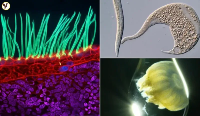
The winning video, announced in mid-September and reposted to Nikon's LinkedIn page the following week, appeared to show cilia the hair-like structures that sweep mucus out of the airways beating in lung tissue from a child with primary ciliary dyskinesia, a rare genetic disease in which those cilia do not work properly. The sample was magnified around 100 times. Beneath the cilia sat red, blue and purple structures.
Within days, microscopy researchers were raising objections in public.
Edward Phelps, a bioengineering researcher at the University of Florida in Gainesville, went through the structures one by one. The purple objects resembled mitochondria — but a sub-epithelial structure made of extracellular mitochondria the size of nuclei does not occur in biology. The blue nodules looked like nuclei and did not behave like nuclei. The red structures he could not identify at all. Commenting on Nikon's LinkedIn post, he wrote that the nuclei "do not behave as nuclei" and that the cilia "appear from nowhere."
Nikon has since deleted that post.
Melanie White, a developmental biologist at the University of Queensland, put the underlying principle to Nature: scientific images are data, and they have to be grounded in the measurement they came from. Andrew Moore, who has previously judged the competition, told The Telegraph the entry reminded him of "old photographs restored with AI." Patrick Hickey, who placed fifth, said the rules plainly prohibit generative AI and require images to come from a microscope.
The argument is not whether AI was used. It is where the line falls
This is the part most coverage flattens, and it is the genuinely interesting question.
Ning Xu, the optical engineering researcher behind the entry, does not deny that a neural network was involved. His account is that the original cilia footage and its motion are real, and that AI was applied only to reconstructed greyscale data, to distinguish and colour structures with similar morphology principally to improve the visual presentation. He has also said his team made no anatomical claims about what the coloured features represent.
Nikon's own blog post was updated to describe an unsupervised neural-network method used in post-processing to distinguish and visualise features in greyscale data. Nikon said Xu was cooperating and had provided technical documentation covering his equipment, imaging methods and processing.
The scientific objection is that in a microscopy image, colour is not cosmetic. Assigning colours to structures is a claim about what those structures are. A viewer looking at a purple object beneath an epithelium reads it as an organelle in a location, at a size and if nothing like that exists, the image has asserted something false regardless of whether the words accompanying it made a formal claim. "We did not say what they were" is a weaker defence for a picture than it would be for a sentence, because a picture is understood immediately and without qualification.
That is the boundary this case sits on: visualisation, which clarifies what the data already contains, against generation, which adds what the data does not. It is an argument happening in laboratories and journal offices everywhere right now, and this is the first time it has cost someone a prize in public.
Nikon's ruling, in its own terms
Nikon's finding was that the entry "did not comply with the competition rules regarding generative AI." The company was careful about the scope of that: it said the decision concerns eligibility and is not a judgement on the entrant's professional reputation or his scientific contributions.
The competition judges on originality, informational content, technical proficiency and visual impact. Nikon has said it is reviewing the rules and procedures of a contest that has run for 15 years without a comparable incident.
Nguyen Nam Nhat, a Vietnamese researcher, moves up from second place to first, for a video of a microscopic roundworm encountering a single-celled organism called a Dileptus. The prize is US$3,000.
Why the microscopy community pushed as hard as it did
The open letter circulated by researchers did not frame this as a dispute over a cash prize. It warned that the video could mislead public understanding of primary ciliary dyskinesia, and urged clinicians and researchers to sign and press Nikon to enforce its own rules.
That is a specific and more serious concern. PCD is a real condition affecting real children, and the images that circulate about a rare disease shape how patients, families and even clinicians picture it. A widely shared, prize-endorsed video carrying structures that cannot exist is a problem for that understanding whether or not anyone intended it as one.
Two details still unsettled
Reporting differs on where Xu works. One account places him at the National University of Singapore; another describes him as a Tsinghua University researcher "at the time," which may indicate a previous post rather than a contradiction. His name also appears as both Ning Xu and Nin Xu across outlets. Neither point is central, but neither is confirmed.
What is settled is the shape of the thing. A competition that had gone 15 years without this problem has now had it, the detection came from the scientific community rather than the organisers, and part of the evidence was a watermark the AI left behind on its own.
Questions readers ask
Why was the Nikon Small World in Motion winner disqualified?
Nikon found that the winning entry "did not comply with the competition rules regarding generative AI." The company said the ruling concerns eligibility only, and is not a judgement on the entrant's professional reputation or scientific contributions. The contest requires images to come from a microscope and prohibits generative AI.
How was the AI use detected?
Partly by a watermark. Ian Donovan, a PhD student at UT Southwestern Medical Center, identified a SynthID watermark in the footage — the AI watermarking technology pioneered by Google DeepMind, which embeds a machine-readable signature in AI-generated content. Separately, microscopy researchers publicly questioned the biological plausibility of structures in the video within days of the result.
What did scientists say was wrong with the video?
Edward Phelps, a bioengineering researcher at the University of Florida, said the purple structures resembled mitochondria but that a sub-epithelial structure made of extracellular mitochondria the size of nuclei does not occur in biology. He said the blue nodules looked like nuclei without behaving like them, and that he could not identify the red structures at all. Commenting on Nikon's LinkedIn post, he wrote that the nuclei "do not behave as nuclei" and the cilia "appear from nowhere."
What is the entrant's explanation?
Ning Xu says the original cilia footage and its motion are real, and that AI was applied only to reconstructed greyscale data in order to distinguish and colour structures of similar morphology, mainly to improve the visual presentation. He says his team made no anatomical claims about what the coloured features represent. Nikon said he cooperated and supplied technical documentation on his equipment, imaging methods and processing.
Who has won the competition now?
Nguyen Nam Nhat, a Vietnamese researcher, moves from second place to first for a video of a microscopic roundworm encountering a single-celled organism called a Dileptus. The prize is US$3,000. Nikon says it is reviewing the rules and procedures of the competition, which has run for 15 years without a comparable incident.